Congenital Midline Cervical Cleft – A Case Report
Y.S. Mohamed, M.V. Peiris
Oral and Maxillofacial Surgery Unit, DGH Polonnaruwa
Abstract
Congenital midline cervical cleft is a rare developmental anomaly of the anterior neck,
mostly found in Caucasian females. Its embryologic origin has not been clearly established,
but the most widely accepted etiology is impaired midline fusion of the distal branchial
arches. When observed at birth the characteristic features are a defect of the ventral area of
the neck with a subcutaneous fibrous cord and a nipple like projection at the upper part. The
defect lies between the mental area superiorly to the suprasternal notch inferiorly with
variable width and length. Associated congenital heart diseases have also been reported.
Treatment is surgical excision and closure of the defect. Since linear scars are more
noticeable than broken lines, closure with z-plasty is done to prevent neck contracture. We
report a case of a 10 month old female infant presented to us at birth with a congenital
midline cervical cleft and a history of an associated atrial septal defect. A z-plasty was carried
out after the excision of the lesion and satisfactory aesthetic and functional results were
achieved. Congenital midline cervical cleft is sometimes misdiagnosed as a thyroglossal duct
anomaly, dermoid cyst, branchial cleft anomaly or birthmark. However, early diagnosis and
suitable surgical treatment are essential to avoid changes in facial development and limitation
of neck extension.
Keywords: Congenital midline cervical cleft. Congenital Anomaly. Branchial
region/abnormalities. Neck/surgery. Reconstructive surgical procedures.
Introduction
Congenital midline cervical cleft (CMCC) also known as median fissure of the neck, web
neck or pterygium colli medianum, is a deformity found at the midline of the neck. It is a rare
developmental anomaly with less than 100 cases reported in literature. Its embryologic origin
has not been clearly established, but most authors classify it as a developmental abnormality
of the branchial arches [1]. When observed at birth the characteristic features are a defect of
the ventral area of the neck with a subcutaneous fibrous cord and a nipple like projection at
the upper part. Congenital midline cervical cleft may be associated with other anomalies like
thyroglossal duct cyst, ectopic bronchogenic cyst, dermoid cyst and may be associated with
congenital heart diseases. Although developmentally related to these disorders, the congenital
midline cervical cleft represents a distinct anomaly that should be recognized at initial
examination The treatment of CMCC consists of complete resection of the lesion followed by
primary closure using W-plasty or, most commonly, single or multiple Z-plasty.
Case Report
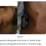
We report a case of a female infant with a congenital midline cervical cleft and a history of an
associated atrial septal defect. This child was referred to the Oral and Maxillofacial Surgery
Unit, DGH Polonnaruwa soon after birth. The infant however had no family history of
congenital defects and her parents were also in good health. On examination she was found to
have a midline vertical skin thickening extending from the mental area superiorly to the
suprasternal notch inferiorly with a fistula like appearance at the upper part. However no
discharges were present from the lesion.
An Ultrasound Scan was taken at the age of 3 months. It was normal and no thyroglossal duct
cyst was evident. A CT scan was obtained at 5 months of age. It revealed an anterior cervical
skin thickening (maximum thickness being 2.8mm) which is 35mm long and extending
inferiorly from the region of the symphysis menti. Thyroid gland was revealed to be normal.
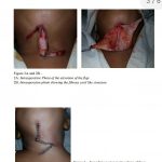
A simple z-plasty was carried out at the age of 10 months after obtaining the consent of the
parents. The fibrotic lesion was completely excised. The skin defect was closed by z-plasties.
Dermal sutures were first placed with 3/0 vicryl and skin was sutured using 5/0 prolene.
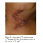
Post-operative period was uneventful and there was no wound infection. Satisfactory
aesthetic and functional results were achieved. In a follow-up examination, 1 and 2 weeks
after the surgery, there was satisfactory wound healing of the Z-plasty and no wound
contracture in the neck was seen.
Discussion
Congenital midline cervical cleft is a rare developmental abnormality of the anterior part of
the neck as first recorded by Luschka in 1848 and noted in the English literature by Bailey in
1924 [2, 3]. However it was completely described by Ombreadanne in 1946. The exact
incidence is unknown, but comprises up to 2% of all neck anomalies and appears to be more
common in Caucasian females [4]. The embryologic mechanism of this anomaly is not yet
clear. In normal embryogenesis the branchial arches grow medially and merge cephalad to
caudal. Prior to fusion, mesodermal tissue migrates between the arches and pushes ectoderm
outward to flatten the ventral furrow [4, 5]. However, the most accepted embryologic
mechanism for the development of this anomaly is impaired fusion of the first or, more
commonly, second branchial arches in the midline as well as improper interaction between
the ectoderm and the mesoderm [6]. Diagnosis of the lesion is by clinical examination and the
anomaly presents as a defect of the ventral area of the neck with a subcutaneous fibrous cord
and a nipple like projection at the upper part. If not treated in newborns, the midline cord
begins to act as a tether as the infant grows. Therefore, surgical excision has both cosmetic
and functional benefits. Congenital midline cervical cleft may be associated with other
anomalies like thyroglossal duct cyst[7, 8], ectopic bronchogenic cyst[9, 10], cleft lower lip,
tongue and mandible[11], cleft sternum, and ectopia cordis with intracardiac anomalies[12].
Associated congenital heart diseases have been reported occasionally with cleft sternum or
ectopia, too [13]. The treatment for congenital midline cervical cleft involves a surgical procedure. Z-plasty is one of the surgical procedures used to correct a congenital midline
cervical cleft and is a simple and routine reconstructive plastic surgical technique. It allows
the surgeon to elongate a contracted scar, reorient the direction of a scar or defect, rotate the
scar tension line, and improve soft tissue contour. Because of these characteristics, single,
double, or multiple Z-plasty is the treatment of choice for congenital midline cervical cleft
[13]. Z-plasty involves the rotation of 2 triangular flaps to close a central defect. The classic
Z-plasty is symmetrically designed so that the lateral limbs of the Z are equal to the defect
that needs reconstruction (central limb), and the angles between the lateral limbs and the
central limb are 60°. When the optimal angle is 60°, it allows a theoretical gain in length of
75% and reorients the direction of the central wound by 90° [13]. In this case we have used a
basic single z-plasty as the treatment choice.
Conclusions: Congenital midline cervical cleft is sometimes misdiagnosed as a thyroglossal
duct anomaly, dermoid cyst, branchial cleft anomaly or birthmark. It is important for the
clinician to arrive at a proper diagnosis at the early stages of life, preferably at birth. If left
untreated congenital midline cervical clefts can cause contracture of the neck [14] and
changes in the mandible [5], including limited neck movement [15]. Therefore, early
diagnosis and suitable early surgical treatment are essential to avoid changes in facial
development and limitation of neck extension.
References
1. 3. Kawar B, Siplovich L. Congenital midline cervical skin bridge: a case report. J
Pediatr Surg. 2008;43(3):544-5
2. Luschka H (1848) Veber fistula colli congenital. Arch. Physiol. Heil 7: 25.
3. Bailey H (1924) Thyroglossal cysts and fistulae. Br J Surg 12: 579-589.
4. Agag R, Sacks J, Silver L. Congenital midline cervical cleft. Cleft Palate Craniofac J.
2007;44(1):98–101.
5. Kakodkar K, Patel S, Maddalozzo J. Congenital Midline Cervical Cleft. [Accessed on
August 5, 2014];Otolaryngology.
6. Van der Staak F, Pruszczynski M, Severijnen R, Van de Kaa CA, Festen C. The
midline cervical cleft. Journal of pediatric surgery. 1991;26(12):1391–1393.
7. Maneshka RJ. Congenital midline cervical cleft with a possible thyro-glossal cyst. Br
J Plast Surg. 1961;14:32.
8. Maschka DA, Clemons JE, Janis JF. Congenital midline cervical cleft: case report and
review. Ann Otol Rhino Laryngol. 1995;104(10 Pt 1):808–11.
9. Davis AD. Medial cleft of lower lip and mandible. Plast Reconstr Surg. 1950;6(1):62.
10. Srinivas V, Pang K, Hallam L, et al. Congenital midline cervical cleft with an
underlying bronchogenic like cyst. Pediatr Surg Int. 2009;25(9):811–3.